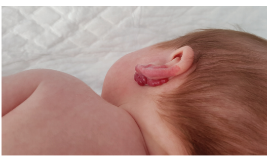
Insights on ulcerated infantile hemangiomas in the ß-blocker era
With Esteban Fernández-Faith, MD

“Although ß-blockers have been standard therapy for problematic infantile hemangiomas (IHs) for a number of years, ulceration continues to be a challenge in many patients with IHs.
Large studies of IH ulceration have not been performed since we started using ß-blockers,” said Esteban Fernández-Faith, MD, Associate Professor of Pediatrics and Dermatology, Ohio State University, and Program Director of the Pediatric Dermatology Fellowship, Nationwide Children’s Hospital, Columbus, Ohio.
To address this gap, gain an understanding about the efficacy of available treatments for ulcerated IH during the “ß-blocker era,” and insights on other characteristics of ulcerated IH, Dr. Fernández-Faith and collaborators from centers of the Pediatric Dermatology Research Alliance (PeDRA) conducted a retrospective cohort study.1
Their findings have implications for therapeutic decisions and anticipatory guidance discussions.
“Our primary goals were to analyze the efficacy of treatments for ulcerated IH and identify prognostic factors for healing time. The results of our analyses show that some ulcerated IHs take a long time to heal; even some ulcerations develop while a patient is on active treatment with a ß-blocker,” said Dr. Fernández-Faith.

“We found larger IH size was the main factor associated with a prolonged time to heal, and our data suggest that if ulceration occurs in an IH needing systemic therapy, a lower dose of propranolol leads to shorter heal times.”
The study included 436 consecutive patients with an ulcerated IH at 1 of 8 PeDRA centers between 2012 and 2016. A total of 364 patients had appropriate data for the analyses of healing time.
The investigation of treatment intervention efficacy classified patients into 5 groups:
1. Wound care only
2. Topical timolol ± wound care
3. Systemic ß-blocker ± wound care
4. Multimodal using at least 2 of the following: topical timolol, systemic ß-blocker, and surgery
5. Pulsed-dye laser (PDL)
With the wound care group serving as the reference, median time to healing was significantly longer in patients treated with multimodal therapy or a systemic ß-blocker and significantly faster in the PDL group.
“We have to consider that this is a retrospective study. Patients were not randomized to treatment and there are probably unaccounted factors that could contribute to the differences observed,” Dr. Fernández-Faith noted. “These findings should not be interpreted to mean that systemic ß-blockers do not work, because they do. Rather, the message is that there are other options that could be used, and the decision should be individualized based on the specific situation.”
A deeper dive into the outcomes of patients treated with systemic ß-blocker therapy showed that the time to healing was two-fold longer among patients treated with a dose of propranolol ≥1mg/kg/day than in those receiving a lower dose.
“To treat IH proliferation, higher doses of propranolol are more efficacious. A dose of propranolol of 2mg/kg/day or higher is commonly used to treat problematic IHs. Based on our research, however, we recommend that if propranolol is indicated for IH treatment and the IH is ulcerated, propranolol should be started at <1mg/kg/day to enable healing and then carefully titrate up to provide the known benefits of propranolol for controlling IH proliferation,” Dr. Fernández-Faith said.
“We can’t explain why the lower dose is better for treating ulceration,” Dr. Fernández-Faith continued. “We need further research to better understand why ulceration happens in the first place.”
Findings for family counseling
Analyses of the effect of IH size on healing time showed median time to heal was 5 weeks for lesions ≤5cm2, 6.14 weeks for those >5 to ≤10cm2, 8 weeks for IHs >10 to ≤50cm2, and 9.29 weeks if the IH was >50 to ≤100cm2. The results showing the importance of IH size can help clinicians with counseling.
“Median time to healing overall was 6.14 weeks, which is quite a long time considering that these lesions cause significant morbidity, and particularly if they are located in the diaper area or around the mouth,” Dr. Fernández-Faith said.
The median age at ulceration development was 13.7 weeks, and as reported in previous research, the vast majority of ulcerations developed in the first 6 months of life. An important minority of the cohort, 17%, developed ulceration after treatment for IH proliferation was started.
“Although we can tell parents or caregivers when ulceration is most likely, the message here is that the possibility for ulceration remains even with treatment and beyond the active proliferation phase of IHs,” Dr. Fernández-Faith emphasized.
Classification system
“One of the main roadblocks we encountered as we set out to conduct this study was to come up with a classification system for IH ulcerations. Looking at previous papers we found researchers used different methods that make it hard to make objective comparisons,” Dr. Fernández-Faith said.
“Our method was based on objective analysis of ulceration in photographs,” Dr. Fernández-Faith noted. “We hope other researchers will find it useful. There is a need for a standardized classification system for IH ulceration severity.”
The classification system includes a subgroup of aggressive ulcerations. “Aggressive ulcerations are very challenging to treat and accounted for approximately 10% of the lesions in our study cohort,” Dr. Fernández-Faith said. “The aggressive ulcerations can cause significant pain, scarring, and seem to get worse no matter what treatment is used.
“We are looking at the characteristics of these lesions in more detail now to recognize at-risk clinical features and eventually identify better therapeutic approaches.”
By Cheryl Guttman Krader
REFERENCE
1. Fernández-Faith E, Shah S, Witman PM, et al. Clinical features, prognostic factors, and treatment interventions for ulceration in patients with infantile hemangioma. JAMA Dermatol. 2021;157(5):566-572.

