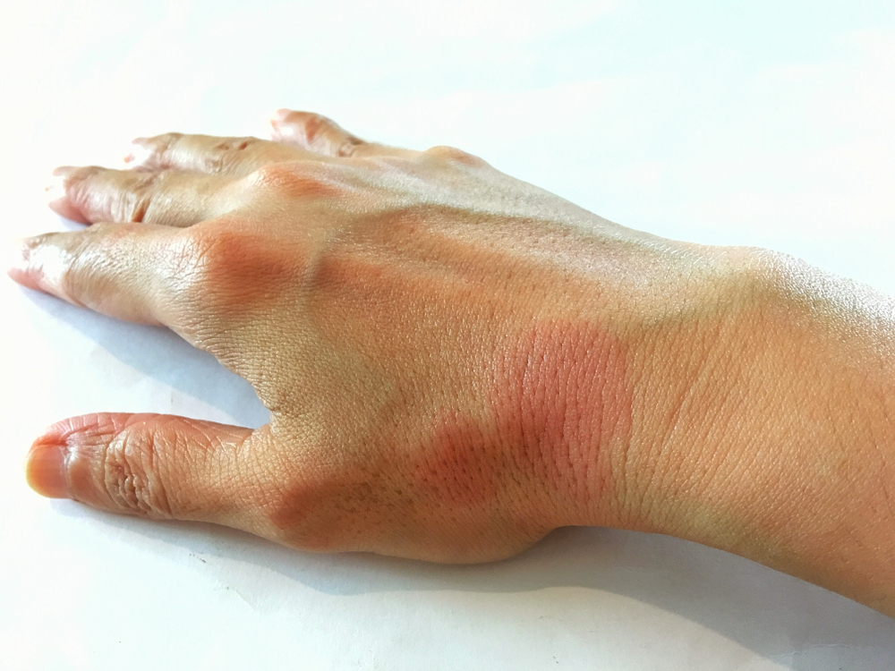
Dr. Jesús Alberto Cárdenas-de la Garza discusses key findings from a recent study identifying the distinctive characteristics of lupus erythematosus tumidus that distinguish it from chronic cutaneous lupus erythematosus.

Jesús Alberto Cárdenas-de la Garza, MD
Professor-Researcher, Department of Rheumatology, Universidad Autόnoma de Nuevo Leόn, Monterrey, Mexico
“Key findings from a retrospective review of patients with lupus erythematosus tumidus (LET) show that it is very rarely associated with the severe manifestations of systemic lupus erythematosus (SLE) and responds well to treatment with hydroxychloroquine in the majority of patients. Therefore, patients diagnosed with LET can be reassured their disease frequently follows a milder course than other subtypes of cutaneous lupus,” said Jesús Alberto Cárdenas-de la Garza, MD, co-author of the recently published study.1
Dr. Cárdenas-de la Garza and colleagues undertook the project recognizing the paucity of information about LET.
“Although it was first described in 1909, most information about LET comes from case reports and small case series. We were interested in clarifying the demographics, clinical characteristics, and autoantibody profiles of affected individuals, associations between LET and systemic disease, optimal therapy, and prognosis,” he said.
The review included adults ages 18 to 75 years with clinical and histologically confirmed LET who presented to the Department of Dermatology, Wake Forest University School of Medicine, Winston-Salem, North Carolina, between July 2012 and July 2018. A total of 25 patients were identified. Their mean age at diagnosis was 46 years, which is slightly older than the typical age of onset for SLE. Eighty-eight percent of patients were non-Hispanic Whites, and the remaining 12% were Black. In contrast to other published series, females predominated over males (4:1).
Lesions most frequently involved the trunk (72%) and upper extremities (72%). Sixty percent of patients had head and neck involvement, and the lower extremities were affected in one-third of patients. Plaques (72%) and papules (68%) were the most common morphologies. The majority of patients reported painful lesions (56%), pruritus (52%), and photosensitivity (52%).
Approximately two-thirds of patients experienced well-defined flares of disease activity. Only 16% of patients were deemed to have concurrent SLE, and single patients each had lupus nephritis and discoid lupus erythematosus.
Only 23% of patients tested for autoantibodies were antinuclear antibody (ANA) positive with a titer >1:80. In addition, three patients had an ANA titer of 1:80 or 1:40.
“These latter low titers are relatively common in healthy women,” Dr. Cárdenas de-la Garza said.
Anti-Ro and anti-La antibodies were each found in one patient, and two had positive anti-double-stranded DNA antibodies.
The most commonly prescribed treatments were topical corticosteroids (84%), oral hydroxychloroquine (80%), and topical calcineurin inhibitors (40%).
“Sixty percent of patients treated with hydroxychloroquine achieved clear or almost clear status, and most were kept on the medication indefinitely. Patients who stopped hydroxychloroquine after a period of 6 to 12 months without flares frequently experienced disease recurrence,” said Dr. Cárdenas-de la Garza.
“However, the retrospective design of our study prevents us from making recommendations about treatment duration or drawing conclusions about relapse rates.”
Patients who failed hydroxychloroquine went on to quinacrine or thalidomide, usually as combination therapy. All patients benefited while on thalidomide, including three of four patients who achieved complete clearance, and thalidomide was generally well tolerated.
“One patient experienced peripheral edema that required stopping the treatment. Thalidomide was not interrupted in another patient who developed gastrointestinal symptoms. None of the patients treated with thalidomide developed peripheral neuropathy,” said Dr. Cárdenas-de la Garza.
Four patients required brief courses of systemic steroids during severe flares. Other treatments included quinacrine (16%), methotrexate (16%), dapsone (4%), and clofazimine (4%).
Classification Conundrum
Originally considered a form of chronic cutaneous lupus erythematosus (CLE), LET was reclassified in 2009 into a new subtype of CLE – intermittent CLE. Whether it should be considered a subset of SLE or represents an entirely separate disorder remains controversial.
“The unique clinical course of LET and its very rare association with systemic lupus raises the question of whether it should be classified in the lupus erythematosus-specific skin disease spectrum. Based on our current understanding, I believe that the classification of LET in its own category of intermittent CLE versus in the chronic CLE group alongside discoid lupus and lupus profundus is justified considering that LET has distinctive characteristics,” said Dr. Cárdenas de-la Garza.
“I also believe that as evidence accumulates, the classification of LET will evolve such that it will not be included in the SLE or CLE disease spectrum. However, any change in classification and the precise classification of LET is not imminent given the scarcity of reported cases.”
Dr. Cárdenas-de la Garza concluded, “There is definitely a need for more research and consensus in LET. Multicenter participation and systematic review of the published series may help bridge current knowledge gaps.”
By Cheryl Guttman Krader
Reference:
1. Pona A, Cardenas-de la Garza JA, Broderick A, et al. Lupus Erythematosus Tumidus Clinical Characteristics and Treatment: A Retrospective Review of 25 Patients. Cutis. 2022;109(6):330-332. doi:10.12788/cutis.0544.

